Dystocia in Laboratory Mice and Rats: Causes and Treatments
When managing your rodent colonies, all stages of the reproductive cycle need to be taken in consideration (see our previous blogs: Breeding Rats and Mice (Part 1): Setting up for Success & Breeding Mice & Rats (Part 2): How to Support Pregnancy and Pup Rearing). Careful planning is required, especially to make sure that your colony management is in alignment with the 3Rs, a framework to perform more humane animal research (1):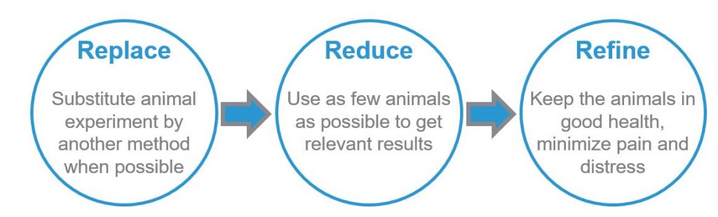
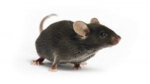
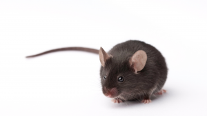
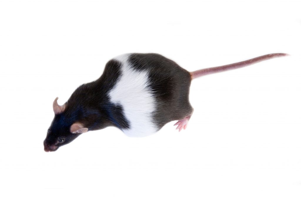
In breeding colonies, pay special attention to the time around birth. Delivery can be painful. One of the most common reproductive-associated conditions in mouse colonies is dystocia. Let’s review this condition, its signs, causes and treatments.
Dystocia Signs and Symptoms
Dystocia comes from Greek “dys” (difficult, painful, disordered) and “tokos” (birth). Dystocia is then defined as difficult, abnormal or dysfunctional labor (2). Mice usually give birth during the night or early morning (dark period in the animal room), and dystocia is thus discovered first thing in the morning, by the animal care technician or veterinarian doing the health check or cage change. Rapid identification and quick response is key to addressing this situation, which if unattended will lead to loss of both female and litter. Training should be provided to recognize the signs of dystocia, that can include:
- Pup visibly stuck in the birth canal or the cervix – The rodents uterus is Y-shaped, with two uterine horns connecting into the cervix. At the point where they meet, a pup could get stuck (3).
- Extended labor that is slow and not progressing – In normal conditions, a pup is delivered about every 15-30 minutes (4). Hard contractions with no results, cessation of contractions or no additional birth within an hour despite the appearance of more pups in the uterus, or labor lasting for more than 2 hours, can all be signs of dystocia.
- Immobility – In normal deliveries, the female will help deliver the pup, and then clean the newborn to clear their airway so oxygenation of the blood can occur, and ingest the placenta for extra protein. If the dam is hunched, weak or limp with little muscle tone, showing poor general condition (5) and presenting clinical signs of distress such as rough hair coat and squinty eyes (6), she can be experiencing dystocia.
- Dehydration – Assess the hydration status with the turgor test, by pinching the nape of the neck; if the skin remains bunched up, the animal is dehydrated (7).
Dystocia Risk Factors and Causes
Dystocia is classified in two categories (8):
- Functional dystocia is associated with factors that are inherent to the dam. Causes can range from uterine inertia (lack of contractions, or contractions that are too weak and infrequent and result in failure to deliver the pups), exhaustion from prolonged contractions or overbreeding, or narrow or non-dilated birth canal (9).
- Obstructive dystocia may result from an oversized fetus that is too large to pass along the maternal birth canal, a fetus that is in the abnormal orientation as it enters the birth canal, or a congenitally malformed or dead fetus (10).
While certain genetically modified mouse strains are more susceptible to dystocia due to gene mutations or knockouts (11-14), some general factors can also put the female at greater risk for dystocia:
- Age. Studies have shown that rate of dystocia was notably higher in older females (≥ 20 weeks) (15-16). Breeders are usually retired at 6-8 months of age for mice and 9-12 months of age for rats.
- Stress. Stress around parturition disrupts birth in many species, including humans, pigs, and rodents (17). Stress can delay parturition in rodents, and can also lead to fetus reabsorption.
- Nutritional deficiency. Hypocalcemia (low calcium) and vitamin E deficiency have been linked to dystocia in different species, such as in goats (18), buffaloes (19), cats or dogs (20).
Check out our nutritional supplement for breeders, DietGel® Prenatal

Request your FREE sample TODAY!
Treatments of Dystocia in Laboratory Mice
When dystocia is discovered, having a standard operating protocol (6) in place for treatment can save time and allow appropriate action to be taken to reduce animal suffering and mortality.
- Removal: A stuck pup can be manually removed by applying a lubricant around the pup, and gently grasping and pulling it with gauze (7). Palpate the abdomen to assess if there are still additional pups to be born.
- Medical management: If the female is still in good health, pursue with a medical treatment often called a “dystocia cocktail” with oxytocin (the strongest known uterotonic drug, from Greek “oxys” (sharp, quick, swift) and “tokos” (birth), although its use has been controversial (21)), calcium gluconate and SQ fluids (do not give IP injections!). Treatment can also include antibiotics (5) and analgesics. Provide a soft diet, such as DietGel® 76A, DietGel® 31M or DietGel® Prenatal, extra nesting materials, a warming pad, keep in a dark and quiet place, and monitor closely.
- Euthanasia: If the female is in poor condition and shows signs of extreme distress, she is unlikely to push out the additional pups and euthanasia is recommended. If pups are valuable, they can be removed by caesarian section and fostered with another female with pups around the same age. For more on fostering, see our pervious blog When Laboratory Mice Can’t be Parents: How to Circumvent Biology
Dystocia in Laboratory Mice: Causes and Treatments – References:
(1) What are the 3Rs?
(2) Health Evaluation of Experimental Laboratory Mice
(3) A Layman’s Guide to Health, Medication Use, Breeding, and Responsible Care of Pet Rats
(4) Mouse models of preterm birth: suggested assessment and reporting guidelines
(5) Common rodents treatments – McGill
(6) Rodent Breeding & Weaning – OLAR Veterinary Staff
(7) FLSC Standard Operating Procedures for Mice in Dystocia
(8) Dystocia and Obstetric Crises: Etiology and incidence
(9) Dystocia in Small Animals – Merck Veterinary Manual
(10) Quick Reference: Improving Mouse Breeding Success – OLC Berkeley
(11) Uterine Dysfunction in Biglycan and Decorin Deficient Mice Leads to Dystocia during Parturition
(12) The Transcription Factor NFIL3 Is Essential for Normal Placental and Embryonic Development but Not for Uterine Natural Killer (UNK) Cell Differentiation in Mice
(13) Absence of p53 and FasL has sexually dimorphic effects on both development and reproduction
(14) Targeted mutation in beta1,4-galactosyltransferase leads to pituitary insufficiency and neonatal lethality
(15) Progressing the care, husbandry and management of ageing mice used in scientific studies
(16) The effects of advanced maternal age on T‐cell subsets at the maternal–fetal interface prior to term labor and in the offspring: a mouse study
(17) The importance of oxytocin mechanisms in the control of mouse parturition
(18) Peri-parturient hypocalcemia in goats: Clinical, hematobiochemical profiles and ultrasonographic measurements of postpartum uterine involution
(19) Vitamin E and selenium supplementation reduces plasma cortisol and oxidative stress in dystocia-affected buffaloes
(20) Reproductive Emergencies
(21) Oxytocin in the Treatment of Dystocia in Mice